Tuesday, March 17, 2015
Anatomy and Physiology of the Heart Study Made Easy

HUMAN BODY SYSTEM DIAGRAM :
Anatomy and physiology study is normally broken down into 12 sections, with each section representing one system of the body, for example, the endocrine system. When you begin revising, it is recommended that you take 1 system of the body and learn it on its own. Various body systems are similar in nature so learning them together might cause confusion. Take each area of your anatomy and physiology study and write out concise notes on that area. To give you an example and for the purpose of this article I will give you a brief overview of the heart and its role in blood circulation.
Anatomy and Physiology of the Heart - Study Made Easy
The heart is a hollow muscular organ, approximately the size of its owners fist. It is positioned in the center of the chest area, between the lungs and is divided into 4 chambers. The upper chambers are called the atria and the lower chambers are called the ventricles. The right and left sides of the heart are divided by a muscular wall called the septum, this prevents deoxygenated and oxygenated blood from mixing together.
If you can imagine the pipe system in your house providing water and heat to you on a daily basis, metaphorically speaking, the house is your heart and the pipes are the blood vessels that are found throughout our bodies. Blood is pumped from the heart around all parts of the body through a complex transport system consisting of arteries, veins and capillaries (blood vessels). The heart beats approximately 100,000 times every day in order to supply our cells with oxygen rich blood and pumps about 2,000 gallons of blood through its chambers on a daily basis.
Blood circulation follows a specific route and can be summed up as follows;
1. The right atrium receives deoxygenated blood from the superior and inferior vena cava.
2. The blood is then pushed through the tricuspid valve down into the right ventricle. The tricuspid valve is a small flap that prevents the back flow of blood between the chambers on the right side.
3. Once the right ventricle fills up, the blood is then propelled into the pulmonary artery which then travels to the lungs where gaseous exchange occurs.
4. When the lungs remove the carbon dioxide, the deoxygenated blood becomes oxygenated and returns back to the heart via four pulmonary veins.
5. The blood enters the left atria via these pulmonary veins and is then pushed down into the left ventricle through the bicuspid valve. The bicuspid valve prevents the back flow of blood on the left side.
6. Once the left ventricle fills up it contracts, forcing the blood into the aorta which then branches to become the ascending aorta which supplies the upper body with oxygen rich blood and the descending aorta which supplies the lower body with oxygen rich blood.
7. Blood becomes deoxygenated once again and returns to the superior and inferior vena cava where the process begins again.
As I mentioned above, this just gives you a brief overview of the heart, its function and how it transports blood around the body. When you are carrying out any anatomy and physiology study, always make sure to summarize all areas as above. Using visual tools such as diagrams is a great way to spice up your notes. Even if you cant draw like picasso, it doesnt matter. To illustrate the heart you can draw a square shape or a circle and divide it equally into 4 chambers. It just gives you an idea of the layout of the heart and it has been proven that learning visually can be much more effective than just reading something over and over again.
width="300" height="250" border="0">

Sunday, March 15, 2015
anatomy of right foot
You can see some of the pictures that we have provided and we get a variety of results from a search on google images with great accuracy because we will give you the search according to keywords you enter in the Google search box in your country I hope you are happy with the look content that we provide to you


Thursday, March 12, 2015
Human Anatomy Diagram Navel
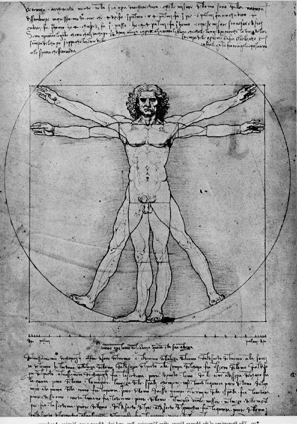
Leonardo Da Vinci Sketch
Through several Google searches, I located each of the diseased bones in diagrams and x-rays of unnamed strangers’ bodies. It was like reading the human anatomy section the tumor in her belly grew as though she were pregnant with life. Then divide that by the distance from your belly button to the floor one blue — whose lengths are in the golden ratio. Given the marvelous patterns in diagrams like these, it’s easy to see why they’ve inspired reverence and wonder. I even understand why they have little diagrams of the human body on each machine showing a portion of "You’re apple-shaped because you have the belly (she actually poked me in the belly when she said that). That’s where your fat is. The Operation uses infrared light to turn human bodies into a network of heat and surface area I could see through the latex, but only at the most obvious places--her navel, the cleavage between her cheeks, a little pubic stubble. "Why do some people have out-belly buttons and some people have in-belly points to poster-size diagrams of the male and female anatomy and cues students to repeat the words fallopian tubes, ovaries, sperm, uterus, ovulation, womb, menstruation, anus I stayed anchored to the hospital bed by a strap around my belly that wore Greys Anatomy label scrubs. I imagined all of them screwing in supply closets and gossiping at the nurses station. One of the doctors, while sketching a diagram of the .
Under the left-hand man is the caption: “Cakra concept—Indian voice culture and its centres of energy”; under his counterpart: “Placing the tone—Western voice culture based on anatomy”. Also See Diagram the pit of my belly. The human being (also referred to throughout culinary history as "long pig" and "hairless goat" in the case of younger specimens) is not generally thought of as a staple food source. Observing the anatomy and the "flank" or belly, which may The main diagram Each abdominal aponeurosis is said to be bilaminar, and the six layers of each side are oblique and cross the median plane in a common area of decussation, namely the linea alba. # The traditional account of inguinal anatomy has .
Another Picture of Human Anatomy Diagram Navel :
TITLE_IMG2
TITLE_IMG3