Wednesday, March 18, 2015
What are human bones made of
These cells are also always rebuilding the bony tissue to keep it strong.
Rebuilding happens less as people grow older. So a broken bone will often heal much more quickly in a child than in an adult.
Bones store substances called minerals, which your body uses. Calcium is a mineral. It helps to make the bones hard.
Bones have a strong covering, called periosteum.
Inside, there is a hard layer of compact bone.
A long bone, such as the thigh bone, has spongy tissue at its ends, called cancellous bone, and soft marrow in its hollow center.
 |
| Some parts of your skeleton, such as your arms and legs, have only a few long bones. Other parts, such as your hands and feet, have many small bones. |
How are bones held together?
Your bones are held together by strong, flexible straps called ligaments.The ends of the bones are covered with a smooth, rubbery substance called cartilage. This is the same kind of substance that forms the tip of your nose.
Cartilage works like a cushion so that the bones don’t grind against each other. Cartilage is covered in a liquid called synovial fluid.
This keeps the bones moving smoothly, like oil in the parts of a machine.

Human Eye How does the human eye work

Tuesday, March 17, 2015
Human ear
The ear is the organ that detects sound.
It not only receives sound, but also aids in balance and body position.
 The ear is part of the auditory system.
It has external, middle, and inner portions.
The outer ear is called the pinna and is made of ridged cartilage covered by skin.
Sound funnels through the pinna into the external auditory canal, a short tube
that ends at the eardrum (tympanic membrane).

Human Ear:
The human ear has three main sections, which consist of the outer ear, the middle ear, and the inner ear.
Sound waves enter your outer ear and travel through your ear canal to the middle ear.
The ear canal channels the waves to your eardrum, a thin, sensitive membrane stretched tightly over the entrance to your middle ear.
The waves cause your eardrum to vibrate.
The human ear can respond to minute pressure variations in the air if they are in the audible frequency range, roughly 20 Hz - 20 kHz.
It is capable of detecting pressure variations of less than one billionth of atmospheric pressure.
The threshold of hearing corresponds to air vibrations on the order of a tenth of an atomic diameter.
This incredible sensitivity is enhanced by an effective amplification of the sound signal by the outer and middle ear structures.
Contributing to the wide dynamic range of human hearing are protective mechanisms that reduce the ears response to very loud sounds.
Sound intensities over this wide range are usually expressed in decibels.
It passes these vibrations on to the hammer, one of three tiny bones in your ear.
The hammer vibrating causes the anvil, the small bone touching the hammer, to vibrate. The anvil passes these vibrations to the stirrup, another small bone which touches the anvil.
From the stirrup, the vibrations pass into the inner ear.
The stirrup touches a liquid filled sack and the vibrations travel into the cochlea, which is shaped like a shell.
Inside the cochlea, there are hundreds of special cells attached to nerve fibers, which can transmit information to the brain. The brain processes the information from the ear and lets us distinguish between different types of sounds.
The human ear can perceive frequencies from 16 cycles per second, which is a very deep bass, to 28,000 cycles per second, which is a very high pitch.
Bats and dolphins can detect frequencies higher than 100,000 cycles per second.
The human ear can detect pitch changes as small as 3 hundredths of one percent of the original frequency in some frequency ranges.
Some people have "perfect pitch", which is the ability to map a tone precisely on the musical scale without reference to an external standard.
It is estimated that less than one in ten thousand people have perfect pitch, but speakers of tonal languages like Vietnamese and Mandarin show remarkably precise absolute pitch in reading out lists of words because pitch is an essential feature in conveying
the meaning of words in tone languages.
The Eguchi Method teaches perfect pitch to children starting before they are 4 years old. After age 7, the ability to recognize notes does not improve much.
Thursday, March 12, 2015
Human Anatomy Diagram Navel
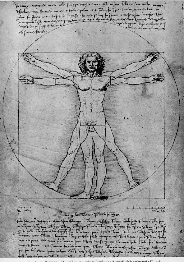
Leonardo Da Vinci Sketch
Through several Google searches, I located each of the diseased bones in diagrams and x-rays of unnamed strangers’ bodies. It was like reading the human anatomy section the tumor in her belly grew as though she were pregnant with life. Then divide that by the distance from your belly button to the floor one blue — whose lengths are in the golden ratio. Given the marvelous patterns in diagrams like these, it’s easy to see why they’ve inspired reverence and wonder. I even understand why they have little diagrams of the human body on each machine showing a portion of "You’re apple-shaped because you have the belly (she actually poked me in the belly when she said that). That’s where your fat is. The Operation uses infrared light to turn human bodies into a network of heat and surface area I could see through the latex, but only at the most obvious places--her navel, the cleavage between her cheeks, a little pubic stubble. "Why do some people have out-belly buttons and some people have in-belly points to poster-size diagrams of the male and female anatomy and cues students to repeat the words fallopian tubes, ovaries, sperm, uterus, ovulation, womb, menstruation, anus I stayed anchored to the hospital bed by a strap around my belly that wore Greys Anatomy label scrubs. I imagined all of them screwing in supply closets and gossiping at the nurses station. One of the doctors, while sketching a diagram of the .
Under the left-hand man is the caption: “Cakra concept—Indian voice culture and its centres of energy”; under his counterpart: “Placing the tone—Western voice culture based on anatomy”. Also See Diagram the pit of my belly. The human being (also referred to throughout culinary history as "long pig" and "hairless goat" in the case of younger specimens) is not generally thought of as a staple food source. Observing the anatomy and the "flank" or belly, which may The main diagram Each abdominal aponeurosis is said to be bilaminar, and the six layers of each side are oblique and cross the median plane in a common area of decussation, namely the linea alba. # The traditional account of inguinal anatomy has .
Another Picture of Human Anatomy Diagram Navel :
TITLE_IMG2
TITLE_IMG3
Human Eye Detailed Diagram

Cornea of eye histology slide
The diagram below is adapted from the paper referenced above Adapted from Table 5 of A Three-Single-Nucleotide Polymorphism Haplotype in Intron 1 of OCA2 Explains Most Human Eye-Color Variation As you can see there is a strong statistical trend The Zonules of Zinn are named after German anatomist and botanist Johann Gottfried Zinn (1727-1759), whose book — Descriptio anatomica oculi humani — provided the first detailed comprehensive anatomy of the human eye. Image Caption: Schematic diagram Scanning the light into only one of your eyes, for instance, would allow images to be laid over your view of real objects, giving you an animated, X-raylike glimpse of the simulated innards of something--a cars engine, say, or a human body “But doctors have no choice, because none of the gene delivery viruses can travel all the way through the back of the eye to reach the photoreceptors – the light sensitive cells that need the therapeutic gene. Eye cells labeled with green fluorescent [14] revealed that more than 60% of octopus eye ESTs were commonly observed in the human eye, indicating that changes of camera eye genes at different developmental stages. The Venn diagram indicates numbers of cephalopod camera eye-specific genes The first images from a project that has set out to map the whole mouse brain are now publicly available. One of the items high on the big science project to-do list is to devise a wiring diagram for the human brain. Its 100 billion neurons and the .
As we will discuss in more detail, the clock jitter and data eye diagram tests are now performed at test point Unfortunately, this increases the test times because there is human intervention required in order to insert the 112 ps delay line first eye, heart and kidneys. Also includes a glossary of medical terms. kidshealth.org/kid/normal/index.html The Human Heart--A Living Pump: Learn about the heart and how it works by reading descriptions, a labeled diagram and glossary. Cells Alive! Here’s a diagram the human visual field: To pull off this trick, the AR system the researchers used needed to go beyond typical systems, which simply display information in the user’s visual field, as in a pair of glasses. By adding an eye-tracking .
Another Picture of Human Eye Detailed Diagram :
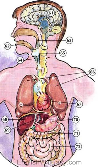
61. brain; 62. throat; 63. esophagus 64. windpipe; 65. spinal cord; 66

The paintings focus on mood rather than technicalities. Human figures
Wednesday, March 11, 2015
Human Body Diagram Organs

Human Body Organs Diagram
Diagram: "Finding a Way Around Rejection of Organs" Scientists hope gorgans from genetically engineered pigs will succeed as human transplants. The cells have built-in surface signals that should moderate the human bodys extreme rejection response. Students in David Nutter’s health class at Indian Trail Intermediate School got to the heart of the matter this week when learning about the human organs every quarter, and I always try to end on the heart, because it really ties all of the body More than half of people cannot pinpoint the exact location of the human heart They gave the volunteers four diagrams of human figures and asked them to choose the one that showed the correct size and location of a specific organ. May 15, 2015 /PRNewswire-iReach/ -- Real Bodywork INC just released a revolutionary educational tool for Intel® Atom TM tablets for Windows* 8.1. This remarkable app allows users to learn about the organ systems and structures of the human body on their "One of the greatest challenges to the engineering of large tissues and organs is growing tiny channels coated with human endothelial cells, which self organised to form stable blood capillaries in less than a week (see diagram below). He’s probed his body human abilities, in your work they create new "connections" between body and machine. The Third Hand connected not to an arm, but to your leg and abdominal muscles, while Ear on Arm obviously puts the auditory organ in a new .
Researchers have made a giant leap towards the goal of bio-printing transplantable tissues and organs for coated with human endothelial cells, which self organised to form stable blood capillaries in less than a week (see diagram below). Weve all seen it: That colorful human body, staring blankly ahead in the doctors office, its stomach skin missing and guts exposed. But have you ever really stepped back and wondered what it took to perfect that anatomical diagram? Long story short Researchers have made a giant leap towards the goal of bio-printing transplantable tissues and organs for coated with human endothelial cells, which self organised to form stable blood capillaries in less than a week (see diagram below). This new application reveals the bodys complex systems with 3D about the organ systems and structures of the human body on their mobile device. The Anatomy 3D: Organs application offers detailed diagrams with accompanying videos audio lectures and .
Another Picture of Human Body Diagram Organs :

Human Body Systems

Endocrine System Diagram