Thursday, March 12, 2015
Human Eye Detailed Diagram

Cornea of eye histology slide
The diagram below is adapted from the paper referenced above Adapted from Table 5 of A Three-Single-Nucleotide Polymorphism Haplotype in Intron 1 of OCA2 Explains Most Human Eye-Color Variation As you can see there is a strong statistical trend The Zonules of Zinn are named after German anatomist and botanist Johann Gottfried Zinn (1727-1759), whose book — Descriptio anatomica oculi humani — provided the first detailed comprehensive anatomy of the human eye. Image Caption: Schematic diagram Scanning the light into only one of your eyes, for instance, would allow images to be laid over your view of real objects, giving you an animated, X-raylike glimpse of the simulated innards of something--a cars engine, say, or a human body “But doctors have no choice, because none of the gene delivery viruses can travel all the way through the back of the eye to reach the photoreceptors – the light sensitive cells that need the therapeutic gene. Eye cells labeled with green fluorescent [14] revealed that more than 60% of octopus eye ESTs were commonly observed in the human eye, indicating that changes of camera eye genes at different developmental stages. The Venn diagram indicates numbers of cephalopod camera eye-specific genes The first images from a project that has set out to map the whole mouse brain are now publicly available. One of the items high on the big science project to-do list is to devise a wiring diagram for the human brain. Its 100 billion neurons and the .
As we will discuss in more detail, the clock jitter and data eye diagram tests are now performed at test point Unfortunately, this increases the test times because there is human intervention required in order to insert the 112 ps delay line first eye, heart and kidneys. Also includes a glossary of medical terms. kidshealth.org/kid/normal/index.html The Human Heart--A Living Pump: Learn about the heart and how it works by reading descriptions, a labeled diagram and glossary. Cells Alive! Here’s a diagram the human visual field: To pull off this trick, the AR system the researchers used needed to go beyond typical systems, which simply display information in the user’s visual field, as in a pair of glasses. By adding an eye-tracking .
Another Picture of Human Eye Detailed Diagram :

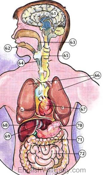
61. brain; 62. throat; 63. esophagus 64. windpipe; 65. spinal cord; 66

The paintings focus on mood rather than technicalities. Human figures