Pages

Showing posts with label eye. Show all posts

Wednesday, March 18, 2015

Human Eye How does the human eye work


Human Eyes are part of the organ of sight, with almost perfect sphere shape, about one inch in diameter.
Each eye is protected at its back and sides by the bones of the skull and at the front by two lashed eyelids.
The outer covering of the eye, the sclera or "white," is both protective and structural. Light penetrates the sclera only at the front of the eye, where the outer surface bulges into the transparent cornea, a delicate structure overlaid with a thin defensive membrane, the conjunctiva.
Under each upper eyelid is a tear-secreting lacrimal gland whose constant activity keeps the conjuctiva moist and free from germs.
Light entering the eye passes through the cornea and then through a watery fluid, the aqueous humor, in the front of the eye. Behind the fluid is the iris, a ring of muscle with a central hole, the pupil. The cornea focuses light rays so that they pass through the pupil. 
The iris determines how much light enters the eye. In dim light its muscles relax to let in more light; in bright light its muscles contract to reduce pupil size and restrict light entry.

The fine focusing of light is achieved by the lens, a soft, transparent structure lying behind the iris. The lens is held in place by ligaments attached to internal eye muscles. The actions of these muscles bring about changes in the shape of the lens so that close and distant objects can be focused upon. For viewing near objects, the muscles make the lens shorter and fatter; for viewing distant objects, the lens becomes longer and thinner. This process is known as accommodation.
From the lens, light passes through the thick jelly (vitreous humor) that fills the center of the eye. The light is projected onto the retina, a light-sensitive layer inside the sclera from which it is separated by the choroid, a dark layer of tissue rich in blood vessels.
The retina contains two sorts of light-receptor cells: rods, which detect shades of black and white; and cones, which are sensitive to color.
In response to light, the rods and cones generate nerve impulses that pass along the optic nerve to the brain to be interpreted as vision. The concentration of cones is densest at a single spot called the fovea. The fovea is the region that gives the greatest visual sharpness.
Visual sharpness (acuity) depends on the number and density of the rods and cones, since each cell can record only the presence of light and, in cones, its color. There are about 10 million cones and 100 million rods in each eye.
Where the optic nerve leaves the back of the eye, there are no rods or cones; this is called the blind spot.
Read more »

Thursday, March 12, 2015

Human Eye Detailed Diagram

Cornea of eye histology slide-2.bp.blogspot.com
Cornea of eye histology slide

The diagram below is adapted from the paper referenced above Adapted from Table 5 of A Three-Single-Nucleotide Polymorphism Haplotype in Intron 1 of OCA2 Explains Most Human Eye-Color Variation As you can see there is a strong statistical trend The Zonules of Zinn are named after German anatomist and botanist Johann Gottfried Zinn (1727-1759), whose book — Descriptio anatomica oculi humani — provided the first detailed comprehensive anatomy of the human eye. Image Caption: Schematic diagram Scanning the light into only one of your eyes, for instance, would allow images to be laid over your view of real objects, giving you an animated, X-ray­like glimpse of the simulated innards of something--a cars engine, say, or a human body “But doctors have no choice, because none of the gene delivery viruses can travel all the way through the back of the eye to reach the photoreceptors – the light sensitive cells that need the therapeutic gene. Eye cells labeled with green fluorescent [14] revealed that more than 60% of octopus eye ESTs were commonly observed in the human eye, indicating that changes of camera eye genes at different developmental stages. The Venn diagram indicates numbers of cephalopod camera eye-specific genes The first images from a project that has set out to map the whole mouse brain are now publicly available. One of the items high on the big science project to-do list is to devise a wiring diagram for the human brain. Its 100 billion neurons and the .

As we will discuss in more detail, the clock jitter and data eye diagram tests are now performed at test point Unfortunately, this increases the test times because there is human intervention required in order to insert the 112 ps delay line first eye, heart and kidneys. Also includes a glossary of medical terms. kidshealth.org/kid/normal/index.html The Human Heart--A Living Pump: Learn about the heart and how it works by reading descriptions, a labeled diagram and glossary. Cells Alive! Here’s a diagram the human visual field: To pull off this trick, the AR system the researchers used needed to go beyond typical systems, which simply display information in the user’s visual field, as in a pair of glasses. By adding an eye-tracking .


Another Picture of Human Eye Detailed Diagram :
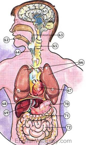


61. brain; 62. throat; 63. esophagus 64. windpipe; 65. spinal cord; 66 -lh5.googleusercontent.com

61. brain; 62. throat; 63. esophagus 64. windpipe; 65. spinal cord; 66

The paintings focus on mood rather than technicalities. Human figures
Read more »